Coinfection of and Leishmania spp. in Synanthropic Reservoirs (Canis familiaris)in an Endemic Area of The State of Quer̮̩̉̉taro, Use of FeSODe as an Antigenic Tool
Villagrán Herrera ME, Valdez FC, Moreno MS, Martínez Ibarra JA and Cabrera JADD
DOI10.21767/2572-5483.100032
Villagrán Herrera ME1*, Valdez FC2, Moreno MS3, Martínez Ibarra JA4 and Cabrera JADD5
1Department of Biomedical Research, Faculty of Medicine, Autonomous University of Querétaro, Santiago de Querétaro, Mexico
2Center for Regional Research, Autonomous University of Yucatán, Mérida Yucatán, Mexico
3Department of Molecular Parasitology, University of Granada, Granada, Spain
4Medical Entomology Area, University Center of the South, University of Guadalajara, Mexico
5Unit of Parasitology and Tropical Medicine, Department of Preventive Medicine and Public Health Autonomous University of Madrid, Spain
- *Corresponding Author:
- María Elena Villagrán Herrera
Department of Biomedical Research
Faculty of Medicine
Autonomous University of Querétaro
Santiago de Querétaro, Mexico
Tel: +524421921200
E-mail: mevh@uaq.mx
Received date: February 22, 2018; Accepted date: March 02, 2018; Published date: March 09, 2018
Citation: Villagrán Herrera ME, Valdez FC, Moreno MS, Martínez Ibarra JA, Cabrera JADD et al. (2018) Coinfection of and Leishmania sp in Synanthropic Reservoirs (Canis familiaris) in an Endemic Area of The State of Querétaro, Use of FeSODe as an Antigenic Tool. J Prev Med Vol.3 No. 2:10.
Copyright: © 2018 Villagrán Herrera ME, et al. This is an open-access article distributed under the terms of the Creative Commons Attribution License, which permits unrestricted use, distribution and reproduction in any medium, provided the original author and source are credited.
Abstract
The importance of synanthropic reservoirs in the epidemiology of metaxenic diseases plays an important role in field studies. Dogs, being the domestic animals that commonly cohabit with the human being in both rural and urban areas, play a fundamental role as reservoirs of blood and tissue protozoa. The migration of the human and its invasion to areas inhabited mainly by natural reservoirs and vectors of Trypanosomatids like Leishmania and Trypanosoma has increased the risk of contracting blood or tissue infection. The present study reveals firstly the seroprevalence of both parasites in canids that coexist openly with the population and in turn are part of the natural fauna of the place (synanthropic reservoir) and secondly the existing co-infection between Leishmania sp in the studied dogs.
Were collected 100 blood samples from canids close to the human and were tested for anti T. cruzi antibodies and anti- Leishmania first with two standard tests, ELISA and Indirect Hemagglutination (HAI) for T. cruzi and Immunochromatographic Test for Leishmania (SD bioline Leishmania Ab), whose results were not convincing. The enzyme superoxydodismutase (SODe) excreted by T. cruzi parasites and Leishmania species present in the endemic region (L. mexicana mexicana, L. infantum chagasi and L. braziliensis braziliensis).
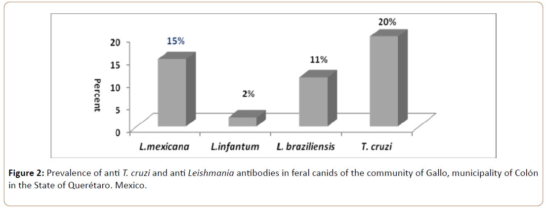
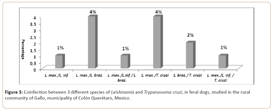
The results obtained reveal a seroprevalence of 20% for T.cruzi, 15% for L. mexicana mexicana, 2% for L. infantum chagasi and 11% for L. braziliensis braziliensis and a coinfection between L. mexicana mexicana/L. braziliensis braziliensis and L. mexicana mexicana/T. cruzi and less frequent, coinfection among L. braziliensis braziliensis/T. cruzi with percentage values of 4%, 4% and 2% respectively.
Keywords
FeSODe; Synanthropic reservoirs; Co-infection in trypanosomatids; Antigenic tool
Abbreviations
HAI: Hemagglutination indirect; ELISA: Enzyme linked immunosorbent assay, SODe: Superoxide diismutase of iron
Introduction
Trypanosomatids are one of the major causes of infections affecting man and animals. Its effects not only cause high rates of morbidity and mortality, but also lead to the production of large economic losses that not only compromise the sick man, but also the family environment and the community in which he is inserted. In some cases they limit social and economic development, as is often the case in many Third World countries [1].
They are protozoan parasites of blood and tissues that affect a wide range of vertebrate hosts. The two most important genera that infect the human and mammals of the world are Leishmania and Trypanosoma that come to produce pathologies of diverse consideration.
Leishmaniasis is a histoparasitosis, produced by protozoans of the genus Leishmania, of intracellular localization (macrophages) that is characterized by presenting cutaneous, mucous or visceral lesions, according to one of the 20 existing pathogenic species and is transmitted through the bite of diptera insects The genres Phlebotomus and Lutzomyia, where there are domestic and wild reservoirs, and it is considered a zoonosis [1]. Distributed in 98 countries around the world, it is considered a cosmopolitan infection. It locates it within the TDR program and indicates as a priority in the research and control programs [2,3].
The WHO estimates an estimated 12 million infected people worldwide and a total of 350 million at risk of infection. In recent years, it has been considered as an emerging disease [4,5], due to climatic changes, population migrations, vector resistance and the difficulty of eliminating animal reservoirs as well as their opportunistic relationship with individuals Immuno compromised (AIDS) [6,7].
With regard to the American Trypanosomiasis better known as Chagas disease, it is a zoonosis of wide complexity that presents endemic only in the Western Hemisphere, from the United States to Argentina. It is produced by the protozoan heterotic Trypanosoma cruzi, a parasite described by Carlos RJ Das Chagas in 1909, who characterized it and established its life cycle by presenting different mechanisms of transmission among which are contaminated feces of triatomine insects of various species (Rhodnius, Pastrongylus, Triatoma and others), blood transfusions, vertical transmission (mother-child), organ transplantation and oral transmission [8]
WHO considers it one of the 9 neglected diseases in the world [3] because it is one of the most complicated parasitic diseases in humans, both because of its unrealistic prevalence and because of its serious clinical picture [9]. In the last 5 years, the investigations revealed that the vectorial transmission reaches 80% of the cases detected and 20% are transmitted by congenital route [10]. It is estimated that 12 million people are infected in 21 Latin American countries [11-13], with 14,000 deaths year [14,15] more than 25 million people are at risk of contracting the disease [13]. Prevalence rates vary widely depending on the geographical area; only in 2008 there were more than 10,000 deaths from Chagas [15].
The impact of the vertical transmission of this disease is estimated for all Latin America in more than 15,000 cases year [16].
Reservoir animals, especially those in close contact with man, play a key role in the transmission of Leishmania and Trypanosoma. The discovery in Brazil in 1913 of the first dog infected by Leishmania [17] raised the possibility of the important role they play in the transmission of the disease. Today, many cases of Leishmaniasis in canines have been described in many countries [18-23], Leishmania mexicana infection was reported for the first time in dogs from the mexican states of campeche, quintana roo and oaxaca [24] which is one of the main links in the peri-urban and urban population due to its sinantropism. The prevalence of Leishmania spp. in dogs from Central and South America varies according to the region and the diagnostic method used with rates from 25% to 75% [25] In Mexico, the prevalence of the disease reaches 58% [26]. Most dogs in rural areas may present cutaneous and/or mucocutaneous lesions, playing a relevant role in the transmission of the domestic cycle of the disease [27,28].
Natural reservoirs in Chagas disease are domestic and wild mammals that play an important role in maintaining the domestic, peri-domestic and wild cycles. Among them, it has been demonstrated that the dog is the fundamental key in the domestic cycle, being considered the overcrowding as a risk factor for the acquisition of the infection in the human beings. This represents a close connection between the parasite's domestic and jungle cycle [29,30]. T. cruzi infection in dogs has been described throughout Latin America, from southern Texas to northern Argentina [31,32].
The diagnosis of both trypanosomatids is very complicated and is usually accompanied by several tests necessary for confirmation. Serological procedures for the detection of antibodies in Leishmania infections are generally based on Indirect Immunofluorescence (IFI) or Immune Enzymatic Assay (EIA) techniques. For cutaneous Leishmaniasis of the Old World seem to have little diagnostic value, instead they are more useful in cutaneous diseases of the New World. The above tests can detect the infection but do not allow identification of the species.
In general, the sensitivity of the tests for skin disease is low. For visceral Leishmaniasis, the direct agglutination test and more recently the development of an rk39 antigen-based indicator test, which together with the PCR, may be specific for genus and species, but are not yet available for application in humans or in animals. On the other hand, the stage of the amastigote can be difficult to detect in the imprints or in the biopsy material and the isolation of the parasite directly or by culture is difficult. In the diagnosis of Trypanosoma cruzi, 3 conventional immunological tests (ELISA, HAI or IFI) are required, two of which must be reactive, so that the confirmed infection is considered, this is considered to be costly, in addition to cross reactions (Leishmania,Toxoplasma, Plasmodium) and obtain a "false positive" result.
The use of detoxifying enzymes as an antigen in the diagnosis of both infections has been very promising in previous studies [33-37]. Other rapid diagnostic techniques such as chromatography and gelatin particles bound to T. cruzi antigen (Serodia) have given results of lower sensitivity.
The study of mixed infections by both trypanosomatids using test with high sensitivity molecular antigens can provide us with data of great interest in the epidemiology of both genera in synanthropic reservoirs.
Materials and Methods
Biological sample
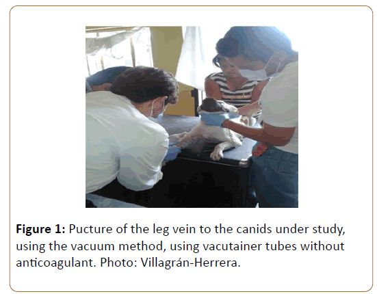
We studied 100 domestic dogs from an endemic area of T.cruzi located in the municipality of Colón in the State of Querétaro. The owners of the animals were asked to sign the consent form, to make the blood sample. At several weekend visits, 100 blood samples of canids were collected by puncture of the central paw vein, in addition to scraping of skin and mucosa (Figure 1).
This protocol was approved by the Bioethics and Research Committee of the Faculty of Medicine of the Autonomous University of Querétaro.
The blood was separated and the serum obtained was frozen in 0.5 ml aliquots in Eppendorf tubes at -30°C for further serological study by the use of molecular antigens excreted by trypanosomatids. Superoxidodismutasa as a defense against free radicals in Trypanosomatids.
The superoxide dismutase (SOD) is responsible for the synthesis of L-arginine by the macrophage against the O2 radical, since Leishmania spp. and Trypanosoma cruzi, do not possess catalase, which disrupts the radical O2 -in H2O2 and O2 both during phagocytosis and in the intracellular phase, in both forms: promastigote and amastigote. H2O2 is converted to H2O by the antioxidant defenses of Leishmania spp.: T (SH) 2 (trypanothione reductase), TXN (trypanoredoxin), PRX (peroxiredoxin) and APX (ascorbate Peroxidase) [38] enzymes involved in the so-called Thiol Redox Balance on which the survival of parasites depends and the spread of infection.
It is possible to consider 4 classes of SOD, which depend on the metallic cofactor present, Cu/Zn-SOD, Mn-SOD and FeSOD. In mammals Cu/Zn-SOD is present at the cytosolic level and Mn- SOD at the mitochondrial level, Cu/Zn-SOD is also present in bacteria whereas Mn-SOD in addition to mitochondrial matrices is also found in prokaryotes. FeSOD is found in prokaryotes, chloroplasts and protozoa. FeSOD and Mn-SOD have very similar sequences and structures so they are believed to have had a common ancestor [38].
Besides being a key key in the proliferation of Leishmania spp. And the fight against ROS (Reactive Oxygen Species), Superoxide Dismutase has been shown to be a good molecular marker in different trypanosomatids: Phytomonas spp. [35,39], T. cruzi [37] and Leishmania L. infantum [39].
FeSOD has been identified and studied in several protozoa and in particular in trypanosomatids more than one isoform has been demonstrated (Phytomonas isolated in Euphorbia characias [39], T. cruzi (Maracay strain) [38] and Trypanosoma brucei.
The extracellular isoform of SOD is considered the most important since it plays a very relevant role in the pathogenesis of many pulmonary, neural and cardiovascular diseases [40,41].
In 2006 Marín et al. Purified Fe-SOD excreted by Phytomonas showing that it had a pI of about 3.6 and a molecular weight of around 22kDa [39,42].
Subsequently, Mateo et al. Purified and characterized FeSOD excreted by T.cruzi, showing that it presented characteristics similar to those found by Marín in 2006, pI of 3.8 and molecular weight in 25kDa [37].
Obtaining FeSODe
Cell culture
For the study and characterization of excreted FeSOD, promastigote forms of Leishmania mexicana, Leishmania infantum chagasi and Lesihmania braziliensis, isolated from dogs and T. cruzi epimastigotes were isolated from an infected patient and cultured in MTL medium (Medium Trypanosomes Liquid) (Gibco®), supplemented with 10% (V/V) fetal bovine serum (SBF, PAAA®), heating to inactivate it at 56°C for 30 minutes.
The growth parasite density (1 × 10⁷cells/ml) was estimated by counting in a Neubauer® chamber and the cells were harvested in the exponential growth phase through centrifugation at 1500 g for 10 minutes at room temperature. The obtained cellular package was resuspended with 25 ml of MTL, without SBF and incubated for 24 hours or overnight at 28°C.
Preparation of fraction H
The promastigotes (Leishmania) and epimastigotes (Trypanosomes), obtained in their exponential phase of growth described in the previous step, undergo a process of lysis or cellular rupture. They are washed twice with phosphate solution pH 7, removing the residues from the culture medium. The pellet is resuspended in 3 ml STE-Buffer 1 lysis buffer (250 nm sucrose, 25 mm Tris-HCl and 1 mm EDTA, pH 7.8), sonicated cold in three cycles of 60 V for 30 seconds (at intervals of 1 min between cycles). The cell lysate was centrifuged (2500 g/10 min/ 4°C), discarding membrane debris, the resulting pellet washed three times with buffer 1 until it had a 9 ml total volume fraction. It was centrifuged the last time in the same previous conditions, obtaining a new supernatant (Fraction H).
Obtaining and purification of excreted FeSOD
The pellet of cells already obtained was resuspended with 25 ml of MTL medium without SBF and incubated 24 hours at 28°C. It is then centrifuged at 1500 xg for 10 minutes, the pellet is discarded and the supernatant is filtered on nitrocellulose membrane (Minisart®). The supernatant is precipitated with ammonium sulfate between 35% and 85%. Again a concentration is made with 35% saline and centrifugation of 9000 g for 20 minutes. The second supernatant is precipitated a second time with ammonium sulfate to a total concentration of 85%. Dissolved the salt is allowed to stand in cold for 20 minutes. Again centrifuged at 9000 xg/20 minutes at 4°C.
The pellet obtained is resuspended in 2.5 ml of distilled water and desalted to 3.5 ml via a Sephadex G-25 (GE Healthcare Life Sciences®, PD 10 column) chromatography column, equilibrated in advance with 25 ml Of distilled water (Fraction P85 or FeSODe-np). The last step is to add 25 μl of antiprotease (Protease Inhibitor Complete Mini, Roche®) thus minimizing the action of the proteases present in the medium.
The P85 fraction of both parasites were purified independently by column chromatography, first by ion exchange, and then by molecular weight filtration.
Once the P85 fraction was concentrated to a volume of 2 ml by lyophilization (LyoQuest, Telstar®), it was passed through an ion exchange column, QAE-Sephadex A-50 (Sigma-Aldrich®), previously equilibrated with the potassium phosphate buffer (20 mm, pH 7.4, 1 mm EDTA), and the elution of the absorbed proteins in the matrix was done through the application of a linear gradient of KCl (0-0.6 M) collecting fractions of 2.5 ml.
The protein concentration of the H P85 and Q1e and Fe-SODe fractions were quantified using the Bradford technique (Sigma Bradford test), using bovine albumin serum as the standard curve.
The obtained FeSODe is used as antigen and the ELISA test is applied for the search for anti-Leishmania and anti Trypanosoma antibodies. [36].
Indirect ELISA Test
With the excreted FeSOD fraction diluted with carbonatebicarbonate buffer (at a final concentration of 1.5 μg and 5 μg), the polystyrene plates are sensitized. 100 μl per well is used. Incubate 2 hours at 37° C or leave overnight in a humid chamber.
To remove unfixed antigens, wash 3 times with 200 μl Wash Buffer solution (Tampon 4; PBS+Twen 20® 0.05%) by overturning the plate on filter paper to dry. Adsorption sites that have been left free because the antigen has not been bound are blocked with buffer 5 (Twen 20®, 0.2% BSA in PBS) and incubated for 2 hours at room temperature and under stirring to prevent binding Not specific between plaque and serum. Wash 3 times again; the plate is incubated for 45 minutes with 100 μl of canine serum diluted 1:80 to be studied.
At the end of the time, the plate is washed 3 times (Buffer 4) and incubated for 30 minutes at 37° C at 37° C and 100 μl of the immunoconjugate (Anti-IgG/anti-dog peroxidase-Sigma-Aldrich) is added at a 1: 1000. Wash again (Buffer 4) and add 100 μl to each substrate well of the immunoconjugate, orthophenylenediamine dihydrochloride (OPD-Sigma-Aldrich®) in Buffer 7; 12.5 ml distilled H2O2, 12.5 ml of 0.1M citratephosphate Buffer, pH 5, Buffer 6 and 10 μl H2O 30%. The plate is incubated in the dark for 20 minutes and 50 μl of reaction stop solution (3 N HCl) is measured at the end of this time by measuring the absorbance at 492 nm in the ELISA reader (Sunrise TM, TECAN). The mean and standard deviation (SD) of the optical density of the negative controls was used for the calculation of the cutoff value (mean + 3x SD).
Results
To evaluate the efficacy of FeSODe from L. mexicana, L.infantum chagassi, L. braziliensis and Trypanosoma cruzi, we tested 100 sera from dogs from the endemic zone of the State of Querétaro, all at a dilution of 1/80 by the technique of ELISA, only 20 were positive to T. cruzi (20%), 15 to (Leishmania mexicana mexicana (15%), 2 to Leishmania infantum chagasi (2%) and 11 to Leishmania braziliensis braziliensis (11%) (Figure 2).
The percentage of coinfection among the Trypanosomatid species were, L. mexicana / L. infantum (1%), L. mexicana/L. braziliensis (4%), L. mexicana/L. infantum/L. braziliensis (1%), L. mexicana/T. cruzi (4%), L. braziliensis/T. cruzi (2%) and for L. mexicana/ L. infantum/T. cruzi (1%) (Figure 3).
Discussion
The diagnosis of Leishmaniasis and Trypanosomiasis is based mainly on the presence of antibodies against both parasites in the serum of infected canines. These antibodies have been detected mainly using different conventional tests such as ELISA, HAI, IFI and Western Blot, as well as rapid tests of immuno chromatography and agglutination in latex particles. In 2002, WHO promotes the ELISA test due to its high sensitivity and low cost, being nowadays one of the main conventional tests.
The problem of using a single test is the presence of crossreactions to parasites belonging to the same or different families, which is why the institution recommends the use of at least two positive conventional serological tests to confirm diagnosis. Different groups of researchers work to improve the quality of these tests based mainly on obtaining antigenic fractions exclusive of the parasite obtained via lysate or excreted by the same to the culture medium. These antigens excreted by parasites of the Trypanosomatid family, such as the FeSOD enzyme, have been proposed as promising diagnostic tools against infection by this group of parasites, examples tested as T. cruzi [33]. Phytomonas [35] and Leishmania spp. [36] Superoxide dismutases are an important group of metalloenzymes that play an important role in the defense of superoxide radicals that protect cells from infection [38,40].
These enzymes are considered to be virulence factors that protect the parasite from attacks by the host cells; their activity was detected in the main Trypanosomatid species [41], in particular the excreted iron superoxydodismutase is the one that has been shown Sensitive in trials with this group of parasites for the T. cruzi species, this enzyme was shown to be highly sensitive and of high diagnostic value in works.
In the present study we show for the first time in a rural community endemic to Trypanosoma cruzi, the importance of domestic canids as reservoirs for the infection of both genera of Trypanosomata, the role of these animals as synanthropic reservoirs to Chagas disease and leishmaniasis is of relevance in this work due to the high prevalence observed for both Leishmania spp and which means that these animals play an important role in the epidemiology of both diseases in the study area.
In 1993, it detected for the first time the presence of leishmaniasis infection in wild dogs in several states of the Mexican Republic [24], mainly in the Yucatán Peninsula, an area endemic for the Leishmaniasis.
The relationship between man and dog so close, both rural and urban, make these animals the first epidemiological link as synanthropic reservoirs [43]. The risk to human infection is greater in terms of the relationship between dogs and their environment as well as the presence of vectors in human dwellings. Different studies support the theory that dogs would be the most important reservoir for Leishmaniasis [18,19,24].
It is therefore necessary to control and supervise such animals thus avoiding a public health problem. The percentage obtained in our study referring to the canine population of Leishmania spp places it among the Latin American countries studied. Our results show the highest frequency of infection in dogs to Leishmania mexicana followed by Leishmania braziliensis and Leishmania infantum chagasi with populations of 15%, 11% and 2%, respectively. The serological prevalence of in these animals was 20%. These data, which are relevant for the first time in the State of Querétaro, demonstrate the importance of these Trypanosomatids in the epidemiology of the respective diseases.
Co-infections observed at both Leishmania spp and T. cruzi were more relevant for Leishmania mexicana and braziliensis and Leishmania mexicana and with Coinfection rates of 4%.
The presence of three species of Leishmania in these reservoirs in the State of Querétaro emphasizes the importance of this endemic until the moment unknown. Regarding, 20% of these animals were seropositive in an area where the level of human infection was 13% [19].
Chagas' disease in Mexico is underestimated according to official data, a prevalence of 1.5% in the Mexican Republic in the only study carried out so far, may lead us to think of a low endemicity for this protozoan, thus the true prevalence This disease is far below reality [44].
It is contradictory that a country with the highest number of vectors counted has so low levels of official endemicity, only in the State of Querétaro the serological prevalence of was 8% in studies carried out by our group [34].
In countries where more than one species of Leishmania is endemic, the differential diagnosis between species becomes indispensable [15]. The two main forms of leishmaniasis found in Mexico are L. mexicana (cutaneous), L. braziliensis (mucocutanea) and L. infantun-chagasi (visceral) [40].
The antigenic fractions of all Leishmania species have been prepared under the same technical conditions; therefore the low prevalence values of L. infantum-chagasi suggest the absence of cross-reaction between different species of Leishmania.The absence of a cross-reaction with Chagas disease is very important because the endemic areas of both diseases are overlapping, which leads to the diagnosis of false positives with consequent aggravating effects on the health of the presumed patient [31].
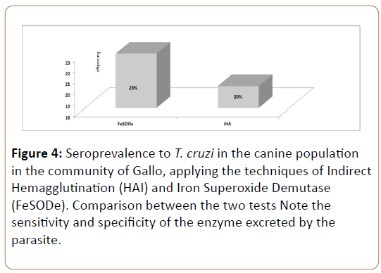
The results presented seem to demonstrate that: the diagnosis performed by the ELISA technique with the antigenic fraction FeSODe is more reliable than by conventional serological methods such as IFI and HAI and that the FeSOD excreted by the 4 parasites did not present cross-reaction between them and other Trypanosomatids such as the case of T. cruzi, thus being able to discriminate between Leishmaniasis and Chagas' disease (Figure 4).
It is for this reason that the results offered in the present work contribute unique findings so far not shown on the importance of the reservoirs synanthropic canines in the epidemiology of leishmaniasis and Trypanosomiasis, with species of Leishmanias not described in our area of study.
Conclusion
L. braziliensis, L. infantum chagasi, L. mexicana and excrete a FeSODe that presents immunogenic characteristics that make it a good molecular marker for serodiagnosis. In conclusion, an indicative ELISA capable of detecting specific antibodies by different species of Leishmania and Tripanosoma has been developed, demonstrating that FeSOD excreted by Leishmania spp. and is an ideal antigen because of its incredible sensitivity and high specificity for the routine diagnosis of these diseases.
Acknowledgements
To the students of the ninth semester of the Faculty of Veterinary Medicine of the Autonomous University of Querétaro, for their support in the blood sampling of the canids.
Disclosure
This research was financed by resources obtained by the PROMEP, from the research professor Autonomous University of Queretaro. There is no conflict of interest.
References
- Atias A (2000) Medical Parasitology. Mediterranean Technical Publications. 1st Edn. 827: 22-264.
- WHO (2010) Control of the leishmaniasis. world health organization technical report series, Geneva 949: 1-186.
- WHO (2007) Neglected diseases: A human rights analysis”.
- Ameen M (2010) Cutaneous leishmaniasis: Advances in disease pathogenesis, diagnostics and therapeutics. Clin Exp Dermatol 35: 699-705.
- Pavli A, Maltezou HC (2010) Leishmaniasis, an emerging infection in travelers. Int J Infect Dis. 14: e 1032-9.
- Gil-Prieto R, Walter S, Alvar J, Gil de Miguel A (2011) Incidence of hospitalizations related to Leishmaniasis by autonomous region and human immunodeficiency virus (HIV) status in Spain. The American Society of Tropical Medicine and Hygiene 85: 820-825.
- Grabmeier PK, Poeppl W, Brunner PM, Rappersberger K, Rieger A, et al. (2012) clinical challenges in the management of leishmania/HIV. coeinfection in a nonendemic area: A case reporte. Case Rep Infect Dis 2012: 1-3.
- Yosida N (2009) Molecular mechnisms of T.cruzi infection by oral route. Mem Inst Oswaldo Cruz 104: 101-107.
- Hotez PJ (2008) Neglected infections of poverty in the USA. PLoS Negl Trop Dis 2: e256.
- Rassi Jr A, Rassi A, Marin-Neto JA (2010) Chagas disease. Lancet 375: 1388-1402.
- Coura JR, Dias JC (2009) Epidemiology, control and surveillance of Chagas disease: 100 years after its Discovery. Mem Inst Oswaldo Cruz 104: 31-40.
- Schummis GA, Yadon ZE (2010) Chagas disease: A Latin American health problem becoming a world health problem. Acta Trop 115: 14-21.
- WHO (2012) Chagas disease (American trypanosomiasis). Descriptive note No 340.
- Coura JRY, Viñas PA (2010) Chagas disease: A new worldwide challenge. Nature 465: S6-7.
- https://www.who.int/wer/2010/wer8534/en/
- https://ghdx.healthdata.org/record/quantitative-estimation-chagas-americas
- Pedrozo AM (1913) Local chaos leishmaniasis. Annals of medicine and surgery 1: 33-39.
- Castro EA, Thomaz SV, Augur C, Luz E (2007) Leishmania (Viannia)braziliensis: Epidemiology of canine cutaneous Leishmaniasis in the State of Paraná (Brazil). Exp Parasitol 117: 13-21.
- Dantas TF (2007) The role of dogs as reservoirs of Leishmania parasites, with emphasis on Leishmania (Leishmania) infantum and Leishmania (Viannia)braziliensis.Vet Parasitol 149: 139-146.
- Dantas-Torres F (2009) Canine Leishmaniasis in South America. Parasit Vectors 2: S1.
- Madeira MF, Schubach A, Schubach TM, Pacheco RS, Oliveira FS, et al. (2006) Mixed infection with Leishmania (Viannia) braziliensis and Leishmania (Leishmania) chagasi in a naturallly infected dog from Rio de Janeiro, Brazil. Trans R Soc Trop Med Hyg 100: 442-445.
- Marco JD, Barroso PA, Calvopiña M, Kumazawa H, Furuya M, et al. (2005) Species assignation of Leishmania from human and canine American tegumentary leishmaniasis cases by multilocus enzyme electrophoresis in North Argentina. Am J Trop Med Hyg 72: 606-611.
- Taranto, NJ, Marinconz,R, Caffaro,CE, Cajal, SP, Malchiodi EL (2000) Mucocutaneous leishmaniasis in naturally infected dogs in Salta, Argentina. Rev Argent Microbiol 32: 129-135.
- Velasco-Castrejón O, Rivas-Sánchez B, Munguía-Saldaña A, Hobart O (2009). Cutaneous leishmaniasis of dogs in Mexico. Infectious diseases and Microbiology. 29: 135-140.
- Cortada VM, Doval ME, Souza Lima MA, Oshiro ET, Meneses CR, et al. (2004) Canine visceral leishmaniosis in Anastácio, Mato Grosso do Sul state. Vet Res Commun 28: 365-374.
- Esquinca RR, Gomez CH, Guevara A (2005) Encuesta rápida de Leishmaniasis visceral en caninos en un área endémica de Chiapas. REDVET 6: 1-7.
- João A1, Pereira MA, Cortes S, Santos-Gomes GM (2006) Canine Leishmaniasis chemotherapy: Dogs clinical condition and risk of Leishmania transmission. J Vet Med A Physiol Pathol Clin Med 53: 540-545.
- Reithinger R, Davies CR (2007) Is the domestic dog ( Canis familiaris) a reservoir host of American cutaneous leishmaniasis? A critical review of the current evidence. Am J Trop Med Hyg 61: 530-541.
- PAHO. Guide for surveillance, prevention, control and clinical management of acute Chagas disease transmitted by food. Para-American Health Organization. Area of ÃÆâÃâââ¬Ãâââ¬Â¹ÃÆâÃâââ¬Ãâââ¬Â¹Health Surveillance and Disease Management. Communicable Disease Project (PAHO / HSD /CD/ 539.09). 2009; Veterinary Public Health Project (Series of technical manuals, 12).
- Cruz-Chan JV, Quijano-Hernandez I, Ramirez-Sierra MJ, Dumonteil E (2010) Dirofilaria immitis and T.cruzi natural co-infection in dogs. Vet J 186: 399-401.
- Rosypal AC, Cortés-Vecino JA, Gennari SM, Dubey JP, Tidwell RR, et al. (2007) Serological survey of Leishmania infantum and T.cruzi in dog from urban areas of Brazil and Colombia. Vet Parasitol 149: 172-177.
- Rosypal AC, Tripp S, Kinlaw C, Sharma RN, Stone D, et al. (2010) Seroprevalence of canine leishmaniasis and American trypanosomiasis in dogs from Grenada, West Indies. J Parasitol 96: 228-229.
- Villagrán ME, Marín C, Rodríguez-Gonzalez I, De Diego JA, Sánchez-Moreno M (2005) Use of an iron superoxide dismutase excreted by T.cruzi in the diagnosis of Chagas disease: seroprevalence in rural zones of the state of Queretaro, Mexico. Am J Trop Med Hyg 73: 510-516.
- Villagrán ME, Sánchez-Moreno M, Marín C, Uribe M, de la Cruz JJ, et al. (2009) Seroprevalence to T.cruzi in rural communities of the state of Querétaro. México: Statistical evaluation of tests. Clinical biochemistry 42: 12-16.
- Marín C, Rodríguez-González I, Sánchez Moreno M (2006) Identification of secreted iron superoxide dismutase for the diagnosis of Phytomonas. Memorias Instituto Oswaldo Cruz 101: 649-654.
- Marín C, Longoni SS, Urbano J, Minaya G, Mateo H, et al. (2009) Enzime-linked inmunosorbent assay for superoxide dismutase-excreted antigen in diagnosis of sylvatic and andean cutaneous leishmaniasis of Peru. Am J Trop Med Hyg 80: 55-60.
- Mateo H, Marín C, Pérez-Cordón G, Sánchez-Moreno M (2008) Purification and biochemical characterization of four iron superoxide dismutases in Trypanosoma cruzi. Mem Inst Oswaldo Cruz 103: 271-276.
- Piacenza L, Irigoín F, Alvarez MN, Peluffo G, Taylor MC et al. (2007) Mitochondrial superoxide radicals mediate programmed cell death in Trypanosoma cruzi: cytoprotective action of mitochondrial iron superoxide dismutase overexpression. Biochem J 403: 323-334.
- Marín C, Hitos AB, Rodríguez-González I, Dollet M, Sánchez-Moreno M (2004) Phytomonas iron superoxide dismutase: a possible molecular marker. FEMS Microbiol Lett 234: 69-74.
- Paramchuk WJ, Ismail SO, Bhatia A, Gedamu L (1997) Cloning, characterization and overexpression of two iron superoxide dismutase cDNAs from Leishmania chagasi: role in pathogenesis. Mol Biochem Parasitol 90: 203-221.
- Ismail SO, Paramchuk W, Skeiky YA, Reed SG, Bhatia A, et al. (1997) Molecular cloning and characterization of two iron superoxide dismutase cDNAs from Trypanosoma cruzi. Mol Biochem Parasitol 86: 187-197.
- Marín C, Sánchez-Moreno M. Excreted /secreted antigens in the diagnosis of Chagas disease. In: Jirillo E, Brandonisio O, editors. Inmune response to parasitic I. Bentham eBooks2010; 10-20 (chapter 2).
- Otranto D, Capelli G, Genchi C (2009) Changing distribution patterns of canine vector borne diseases in Italy: leishmaniosis vs. dirofilariosis. Parasit Vectors 2: S2.
- Petherick A (2010) Country by Country. Nature 465: 10.
Open Access Journals
- Aquaculture & Veterinary Science
- Chemistry & Chemical Sciences
- Clinical Sciences
- Engineering
- General Science
- Genetics & Molecular Biology
- Health Care & Nursing
- Immunology & Microbiology
- Materials Science
- Mathematics & Physics
- Medical Sciences
- Neurology & Psychiatry
- Oncology & Cancer Science
- Pharmaceutical Sciences
